Within 10 Brain
What Brain Scans Do Not Show
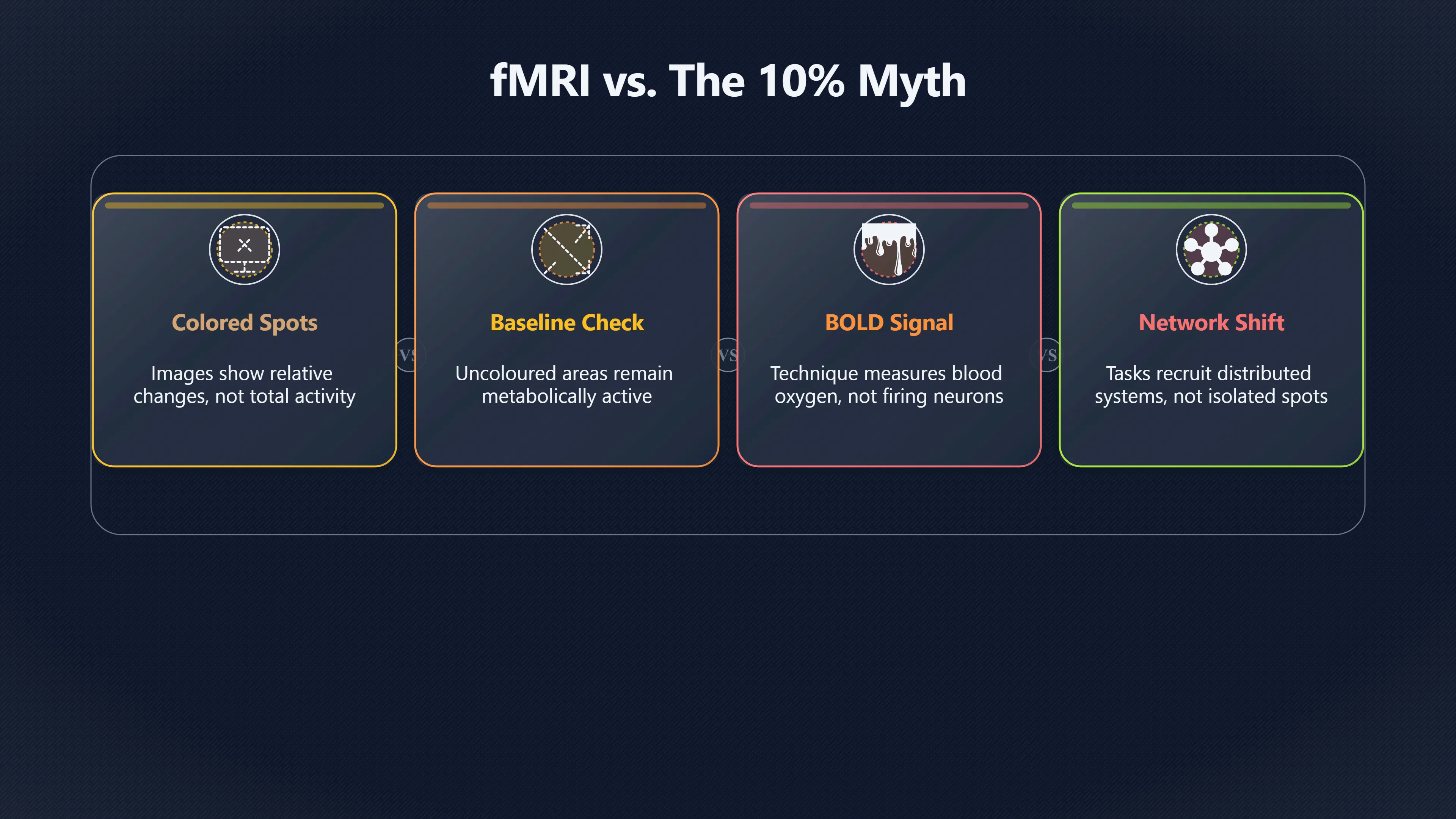
Brain scans highlight relative changes in activity, not vast silent regions waiting to be switched on.
On this page
- Why highlighted scan areas can mislead
- How different tasks recruit different networks
- Why sleep still involves widespread brain activity
Page outline Jump by section
Introduction
Brain scans are often presented as if they reveal exactly which parts of the brain are “on” and which parts are “off”. Bright colours appear on a screen, a few regions light up, and it can look as though most of the brain is sitting idle until a special task activates it. That impression has helped support myths such as the idea that humans use only 10 per cent of their brains.
 Modern brain imaging shows something very different. Techniques such as functional magnetic resonance imaging (fMRI) and positron emission tomography (PET) do not reveal vast inactive regions waiting to be unlocked. Instead, they usually show changes in activity levels across networks that are already functioning. The coloured areas in many published images represent differences relative to a comparison condition, not the only places where the brain is active. PMC [The Open University]open.eduThe Open UniversityHow FMRI works | OpenLearnFunctional magnetic resonance imaging, or FMRI, works by detecting the changes in blood oxyg…
Why highlighted scan areas can mislead
One of the most common misunderstandings comes from the way brain scans are displayed. In many fMRI images, a participant performs a task such as reading words, moving a hand or recognising faces. Researchers then compare brain activity during that task with activity during a baseline condition.
The coloured regions usually mark places where activity increased significantly compared with the baseline. They do not show every active neuron in the brain, nor do they indicate that uncoloured areas are inactive. In fact, the entire brain remains metabolically active throughout the experiment. PMC [The Open University]open.eduThe Open UniversityHow FMRI works | OpenLearnFunctional magnetic resonance imaging, or FMRI, works by detecting the changes in blood oxyg…
Another source of confusion is that fMRI does not directly record neurons firing. The most widely used signal, known as the Blood Oxygen Level Dependent (BOLD) signal, measures changes in blood oxygenation and blood flow associated with neural activity. Researchers infer brain activity from these physiological changes. PMC [Royal Society]royalsociety.orgqa what is boldQ&A: What is BOLD?29 Aug 2016 — Blood Oxygenation Level Dependent (BOLD) imaging is a technique that is commonly used for measuring brain…
This distinction matters because a colourful activation map can look more definitive than it really is. A scan showing strong activation in one region does not mean other regions have stopped working. It usually means that one area changed more than the comparison condition.
The visual style of published images can reinforce the misunderstanding. Researchers often apply statistical thresholds so that only the strongest changes appear in colour. Areas below that threshold may still be active but are not highlighted. The resulting image is designed to show meaningful differences, not to provide a literal picture of the brain switching large sections on and off. [PMC]pmc.ncbi.nlm.nih.govPMCOverview of Functional Magnetic Resonance ImagingPMCby GH Glover · 2011 · Cited by 1859 — Blood Oxygen Level Dependent (BOLD) functional magnetic resonance imaging (fMRI) depicts changes…
How different tasks recruit different networks
Brain imaging has repeatedly shown that mental activity depends on distributed networks rather than isolated hotspots. Different tasks shift the balance of activity across these networks, but they do not awaken previously unused brain territory.
For example:
- Visual tasks increase activity in networks involved in processing visual information.
- Language tasks recruit regions associated with speech, comprehension and memory.
- Attention-demanding tasks engage what researchers often call task-positive networks, involving frontal and parietal regions linked to goal-directed behaviour.
- Internally focused activities such as remembering the past, imagining the future or reflecting on oneself involve parts of the default mode network. Frontiers 3ScienceDirect [The Outside the Box Project]otbproject.com· The…Read more…
The important point is that these networks overlap, interact and constantly exchange information. A person solving a mathematical problem is not using a small isolated section of the brain while the rest lies dormant. Instead, multiple systems coordinate attention, memory, sensory processing, error monitoring and motor control. The pattern changes according to the task. [Deep Blue]deepblue.lib.umich.eduDeep BlueInterplay Between Default-Mode and Task-Positive Networksby C Chen · 2010 · Cited by 1 — Recent work has demonstrated that the h…
Research on the default mode network has been particularly important in correcting simplistic interpretations of brain scans. Earlier studies sometimes treated resting states as a period when the brain was largely inactive. Later work showed that the brain remains highly organised during rest, with distinct networks maintaining ongoing activity linked to self-reflection, memory, social cognition and internal thought. [PMC]pmc.ncbi.nlm.nih.govPMCThe neural basis of the blood-oxygen-level-dependentPMCby NK Logothetis · 2002 · Cited by 1333 — This paper reviews the basic principles of MRI and fMRI, and subsequently discusses in some… [Springer]link.springer.comBrain Activity and Resting State Networks18 Oct 2022 — Resting state functional magnetic resonance imaging (RS-fMRI) has emerged as a maj…
This means that even when someone appears to be doing nothing, the brain is not simply waiting to be activated. It is already engaged in a range of internally directed processes.

Why sleep still involves widespread brain activity
Sleep is another area where brain scans challenge the idea of large unused regions. If the 10 per cent myth were true, sleep might be expected to show long periods of near-total shutdown. Neuroimaging reveals something far more complex.
PET, fMRI and combined EEG-imaging studies show that activity patterns change substantially across sleep stages, but the brain remains active throughout the night. Different regions increase or decrease their activity depending on whether a person is in light sleep, deep non-rapid eye movement (NREM) sleep or rapid eye movement (REM) sleep. [PMC]pmc.ncbi.nlm.nih.govPMCThe Journey of the Default Mode Network: Development…by FR Azarias · 2025 · Cited by 55 — The Default Mode Network (DMN) is a brain… [PMC]pmc.ncbi.nlm.nih.govPMCFunctional Neuroimaging Insights into the Physiology of…by TT Dang-Vu · 2010 · Cited by 377 — PET and block-design fMRI (i.e., cont…
Deep sleep is associated with reductions in activity in some higher-order regions, yet widespread coordinated activity remains present. Researchers observe large-scale oscillations, ongoing communication between brain systems and physiological processes linked to memory consolidation and restoration. [PMC]pmc.ncbi.nlm.nih.govnih.govEEG-fMRI Methods for the Study of Brain Networks during Sleepby JH Duyn · 2012 · Cited by 78 — BOLD fMRI is the most recent neuroi… [Nature]nature.comThe resulting…Read more…
REM sleep provides an even clearer example. During REM periods, several regions involved in perception, emotion and memory can become highly active. Brain activity during REM often resembles wakefulness more closely than popular assumptions about sleep would suggest. [Wiley Online Library]onlinelibrary.wiley.comOnline Library Sleep neuroimaging: Review and future directionsWiley Online LibrarySleep neuroimaging: Review and future directions - Pereira12 Feb 2025 — The results of these studies have shown that…
Recent multimodal imaging studies combining EEG, PET and fMRI have further demonstrated that sleep involves shifting patterns of network activity rather than global inactivity. As sleep deepens, some systems reduce their metabolic demands while others remain dynamically engaged. Nature [Wiley Online Library]onlinelibrary.wiley.comOnline Library Sleep neuroimaging: Review and future directionsWiley Online LibrarySleep neuroimaging: Review and future directions - Pereira12 Feb 2025 — The results of these studies have shown that…
These findings directly contradict the idea that large portions of the brain sit unused for most of the day.
What brain scans actually tell us about the 10 per cent myth
The strongest lesson from modern neuroimaging is not that every brain region is maximally active all the time. Rather, it is that the brain operates through constantly changing patterns of activity distributed across interconnected networks.
Brain scans reveal relative increases and decreases, cooperation between specialised systems and shifts in functional organisation. They do not reveal a vast reserve of silent neural tissue waiting to be switched on. Damage to even small brain regions can produce serious impairments, which is one reason neuroscientists have long rejected the idea that 90 per cent of the brain serves no purpose. Neuroimaging has reinforced that conclusion by showing activity across the brain during work, rest and sleep. PMC [The Open University]open.eduThe Open UniversityHow FMRI works | OpenLearnFunctional magnetic resonance imaging, or FMRI, works by detecting the changes in blood oxyg…
The colourful images that often accompany discussions of the brain are powerful scientific tools, but they are easy to misread. Their real message is not that only a few bright spots matter. It is that the brain is a dynamic system whose activity is constantly being redistributed across networks depending on what a person is seeing, thinking, remembering, feeling or dreaming. [PMC]pmc.ncbi.nlm.nih.govPMCby F Xin · 2015 · Cited by 123 — In contrast to the FPC, the activity of the default mode network (DMN) has been documented as typical… [PMC]pmc.ncbi.nlm.nih.govPMCOverview of Functional Magnetic Resonance ImagingPMCby GH Glover · 2011 · Cited by 1859 — Blood Oxygen Level Dependent (BOLD) functional magnetic resonance imaging (fMRI) depicts changes…

Amazon book picks
Further Reading
Books and field guides related to What Brain Scans Do Not Show. Use these as the next step if you want deeper reading beyond the article.

Livewired
First published 2020. Subjects: Physiology, Internal medicine, Brain, Neuroplasticity, Learning.

The brain
First published 2015. Subjects: SCIENCE / Life Sciences / Biology / General, Brain, Self, SCIENCE / Life Sciences / Neuroscience, Neurosc...

Incognito
First published 2011. Subjects: SCIENCE / Life Sciences / Biology / General, Brain, Subconsciousness, New York Times bestseller, nyt:hard...
eBay marketplace picks
Marketplace Samples
Example marketplace items related to this page. Use the search link to explore similar finds on eBay.


Endnotes
-
Source: pmc.ncbi.nlm.nih.gov
Title: PMCOverview of Functional Magnetic Resonance Imaging
Link: https://pmc.ncbi.nlm.nih.gov/articles/PMC3073717/Source snippet
PMCby GH Glover · 2011 · Cited by 1859 — Blood Oxygen Level Dependent (BOLD) functional magnetic resonance imaging (fMRI) depicts changes...
-
Source: open.edu
Link: https://www.open.edu/openlearn/body-mind/health/health-sciences/how-fmri-worksSource snippet
The Open UniversityHow FMRI works | OpenLearnFunctional magnetic resonance imaging, or FMRI, works by detecting the changes in blood oxyg...
-
Source: pmc.ncbi.nlm.nih.gov
Title: PMCThe neural basis of the blood-oxygen-level-dependent
Link: https://pmc.ncbi.nlm.nih.gov/articles/PMC1693017/Source snippet
PMCby NK Logothetis · 2002 · Cited by 1333 — This paper reviews the basic principles of MRI and fMRI, and subsequently discusses in some...
-
Source: nature.com
Link: https://www.nature.com/articles/s41593-025-02132-9Source snippet
The resulting...Read more...
-
Source: sciencedirect.com
Title: ScienceDirect Task Positive Network
Link: https://www.sciencedirect.com/topics/medicine-and-dentistry/task-positive-networkSource snippet
ScienceDirectTask Positive Network - an overviewThe task positive network (TPN) is defined as a brain network that activates during goal...
-
Source: pmc.ncbi.nlm.nih.gov
Link: https://pmc.ncbi.nlm.nih.gov/articles/PMC12025022/Source snippet
PMCThe Journey of the Default Mode Network: Development...by FR Azarias · 2025 · Cited by 55 — The Default Mode Network (DMN) is a brain...
-
Source: nature.com
Link: https://www.nature.com/articles/s42003-024-06506-wSource snippet
Flexible adaptation of task-positive brain networks predicts...by A Weigard · 2024 · Cited by 12 — Another notable finding concerns the...
-
Source: link.springer.com
Link: https://link.springer.com/rwe/10.1007/978-3-030-88832-9_133Source snippet
Brain Activity and Resting State Networks18 Oct 2022 — Resting state functional magnetic resonance imaging (RS-fMRI) has emerged as a maj...
-
Source: link.springer.com
Link: https://link.springer.com/article/10.1007/s00429-022-02467-0Source snippet
activating the default mode network map multiple...by L Mancuso · 2022 · Cited by 64 — A new deconstructive line of research is pointing...
-
Source: pmc.ncbi.nlm.nih.gov
Link: https://pmc.ncbi.nlm.nih.gov/articles/PMC2982729/Source snippet
PMCFunctional Neuroimaging Insights into the Physiology of...by TT Dang-Vu · 2010 · Cited by 377 — PET and block-design fMRI (i.e., cont...
-
Source: onlinelibrary.wiley.com
Title: Online Library Sleep neuroimaging: Review and future directions
Link: https://onlinelibrary.wiley.com/doi/10.1111/jsr.14462Source snippet
Wiley Online LibrarySleep neuroimaging: Review and future directions - Pereira12 Feb 2025 — The results of these studies have shown that...
-
Source: nature.com
Link: https://www.nature.com/articles/s41467-025-64414-xSource snippet
NatureSimultaneous EEG-PET-MRI identifies temporally coupled...by JE Chen · 2025 · Cited by 11 — Moreover, sympathetic activity that eli...
-
Source: sciencedirect.com
Title: Blood Oxygen Level
Link: https://www.sciencedirect.com/topics/psychology/blood-oxygen-levelSource snippet
an overviewfMRI is based on the principle that brain activity causes a change in the relative amounts of oxy- and deoxy-hemoglobin which...
-
Source: royalsociety.org
Title: qa what is bold
Link: https://royalsociety.org/blog/2016/08/qa-what-is-bold/Source snippet
Q&A: What is BOLD?29 Aug 2016 — Blood Oxygenation Level Dependent (BOLD) imaging is a technique that is commonly used for measuring brain...
-
Source: otbproject.com
Link: https://otbproject.com/brain-networks/Source snippet
· The...Read more...
-
Source: deepblue.lib.umich.edu
Link: https://deepblue.lib.umich.edu/items/5e1036e4-f3b3-4fd2-8de1-be6f92236493Source snippet
Deep BlueInterplay Between Default-Mode and Task-Positive Networksby C Chen · 2010 · Cited by 1 — Recent work has demonstrated that the h...
Additional References
-
Source: facebook.com
Link: https://www.facebook.com/groups/NeuroscienceGroup/posts/10166122945645089/Source snippet
Task-positive and default mode brain networksThe first is the task-positive network, also known as the central executive network. This is...
-
Source: news-medical.net
Link: https://www.news-medical.net/news/20251024/Study-reveals-how-brain-activity-energy-use-and-blood-flow-interact-during-sleep.aspxSource snippet
Study reveals how brain activity, energy use, and blood...24 Oct 2025 — The researchers found that energy use and metabolism decrease as...
-
Source: neupsykey.com
Title: what brain imaging reveals about sleep generation and maintenance
Link: https://neupsykey.com/what-brain-imaging-reveals-about-sleep-generation-and-maintenance/Source snippet
What Brain Imaging Reveals about Sleep Generation and...13 Mar 2017 — It was shown that regional brain activity during sleep was segrega...
-
Source: facebook.com
Link: https://www.facebook.com/thedoctorasky/posts/new-imaging-research-shows-that-the-brain-does-more-than-rest-during-deep-sleep-/1594496135570581/Source snippet
Using simultaneous EEG, PET, and MRI scans, scientists tracked...Read more...
-
Source: Wikipedia
Title: Blood oxygenation level–dependent imaging
Link: https://en.wikipedia.org/wiki/Blood-oxygenation-level%E2%80%93dependent_imagingSource snippet
Blood-oxygenation-level–dependent imagingBlood-oxygenation-level–dependent imaging, or BOLD-contrast imaging, is a method used in func...
-
Source: frontiersin.org
Link: https://www.frontiersin.org/research-topics/404/functional-brain-network-changes-in-human-sleep-in-health-and-disease-the-role-of-neuroimaging/magazineSource snippet
ole of neuroimaging. 123.7K. views. 52. authors.Read more...
-
Source: biorxiv.org
Link: https://www.biorxiv.org/content/10.1101/2021.03.17.435799.fullSource snippet
Recent developments in network neuroscience suggest reconsidering what we thought we knew about the Default Mode Network (DMN).Read more...
-
Source: pmc.ncbi.nlm.nih.gov
Link: https://pmc.ncbi.nlm.nih.gov/articles/PMC3387650/Source snippet
nih.govEEG-fMRI Methods for the Study of Brain Networks during Sleepby JH Duyn · 2012 · Cited by 78 — BOLD fMRI is the most recent neuroi...
-
Source: pmc.ncbi.nlm.nih.gov
Link: https://pmc.ncbi.nlm.nih.gov/articles/PMC4526481/Source snippet
PMCby F Xin · 2015 · Cited by 123 — In contrast to the FPC, the activity of the default mode network (DMN) has been documented as typical...
-
Source: pnas.org
Link: https://www.pnas.org/doi/10.1073/pnas.2016732119Source snippet
fMRI spectral signatures of sleepby C Song · 2022 · Cited by 72 — We show that the fMRI signatures of sleep can be employed to monitor lo...
Topic Tree